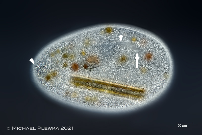
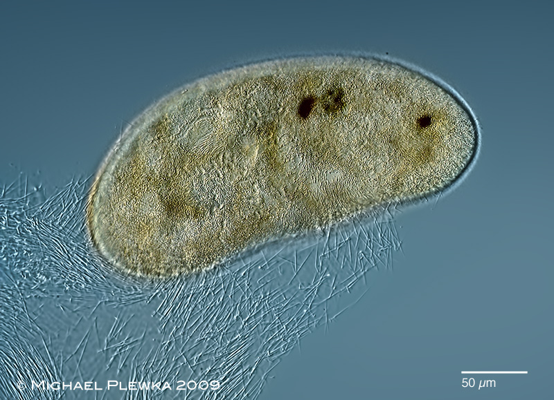
Frontonia leucas: focal plane on the oral apparatus (arrow) and the postoral suture (marked by arrowheads). This specimen has engulfed a diatom. (5)
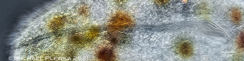
Frontonia leucas: oral apparatus and the postoral suture; detail. (5)
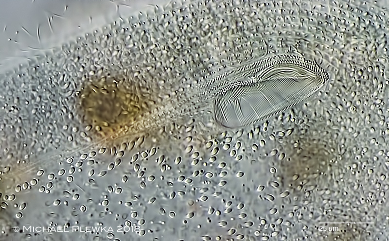
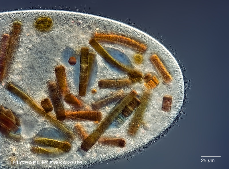
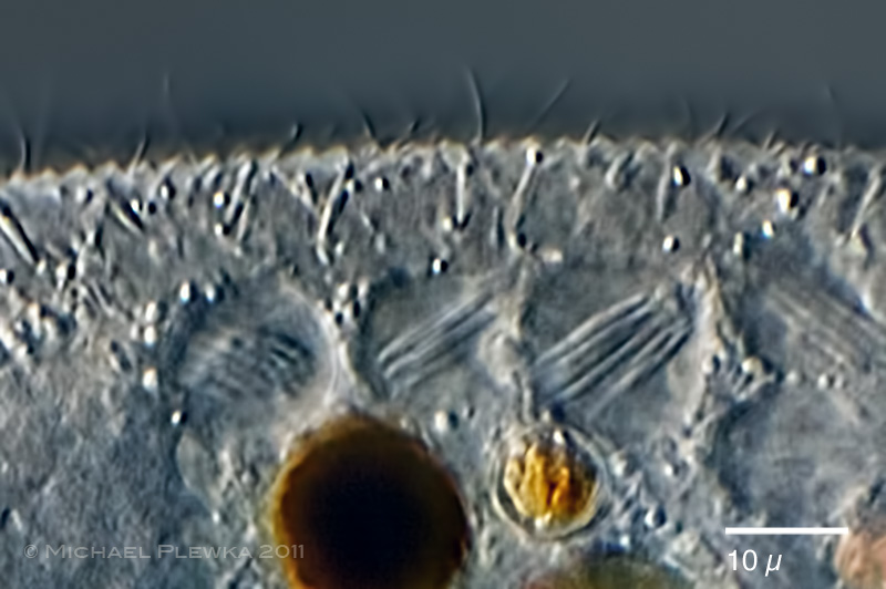
Frontonia leucas: oral apparatus of a specimen from (3) Also visible are the extrusomes (trichocysts) in the cortex.(3)
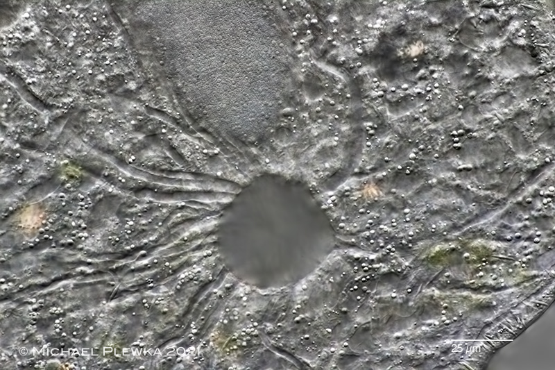
Frontonia leucas: two images of the contractile vacuole (CV). The species is characterized by a single contractile vacuole with long, slightly meandering radial collecting canals, (upper image)(5). Lower image: focal plane on the excretion pore, collecting canals and extrusomes.
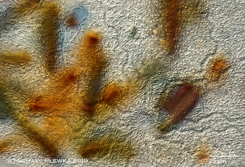
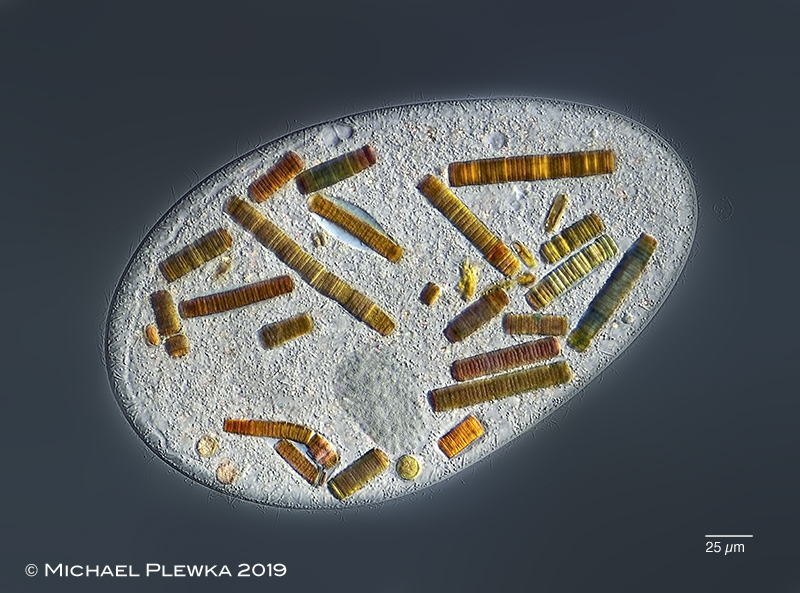
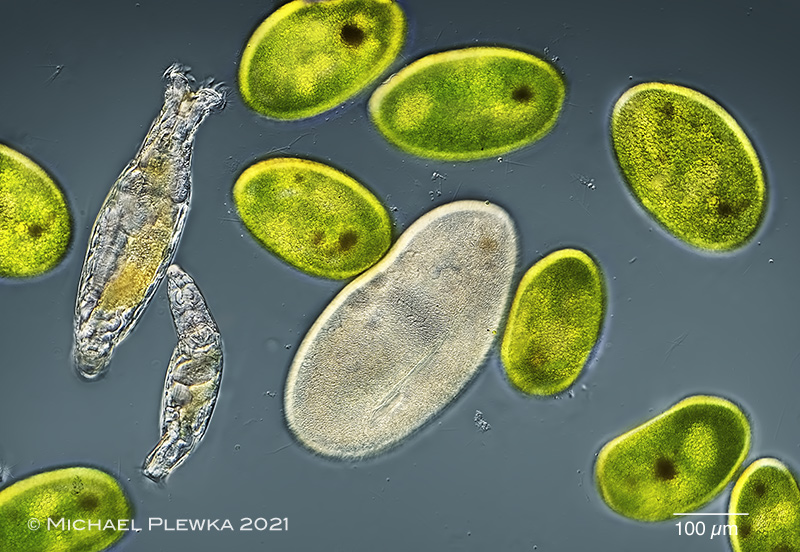
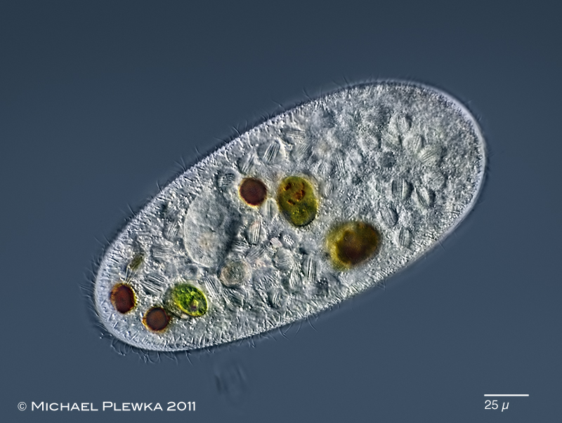
Frontonia leucas: two images of a specimen which has engulfed cyanobacteria. Due to the color pigments that change their color during the digestion process the food vacuoles / cilates become very colorful.
Frontonia leucas: specimen with ejected extrusomes due to a contact with the ciliate Spirostomum sp. (2)
Frontonia leucas: together with the zoochlorellae-bearing Frontonia cf vernalis in location/ habitat No. (4)
Frontonia leucas: specimen infested by the parasitic bacterium Holospora sp. (image below:crop)
Frontonia leucas: Makronukleus und Trichocysten
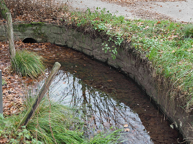
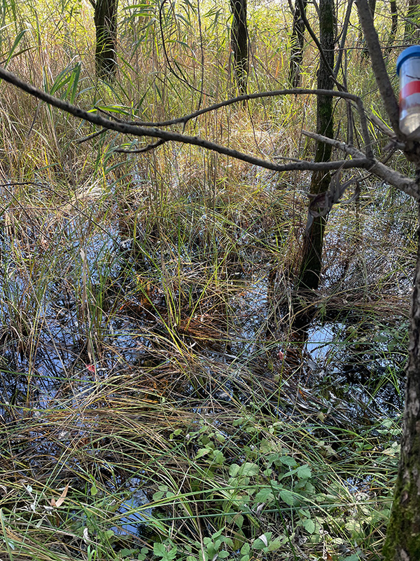
Location (3): Hattingen Wodantal, Heierbergsbach, puddle (click to enlarge)
Habitat (3): detritus
Date (3): 29.11.2018 (1); 10.12.2018 (2); 11.12.2018 (3)